From Equations to Care: Individual Insights That Matter
Grounded in Biology, Guided by Mathematics
From Ion Channels to Organ Systems
A single ion channel switches currents, yet its collective behavior shapes a heartbeat. Ordinary and partial differential equations connect such microscopic actions to organ-scale signals, like blood pressure waves or electrocardiograms. By calibrating these models with imaging and lab values, we relate mechanisms to observations. This bridge explains why two patients with similar symptoms can require very different interventions based on underlying dynamics.
Personalization Through Data Assimilation
Parameter estimation turns general understanding into individual insight. Data assimilation, using Bayesian inference or Kalman filtering, fuses time series, imaging, and labs to tune models for one person. Identifiability analyses prevent overfitting and guide which measurements truly matter. The result is not just a fit curve, but a physiologically plausible state estimate that clarifies causes, predicts responses, and supports timely clinical action with quantified confidence.
Patient Digital Twins in Practice
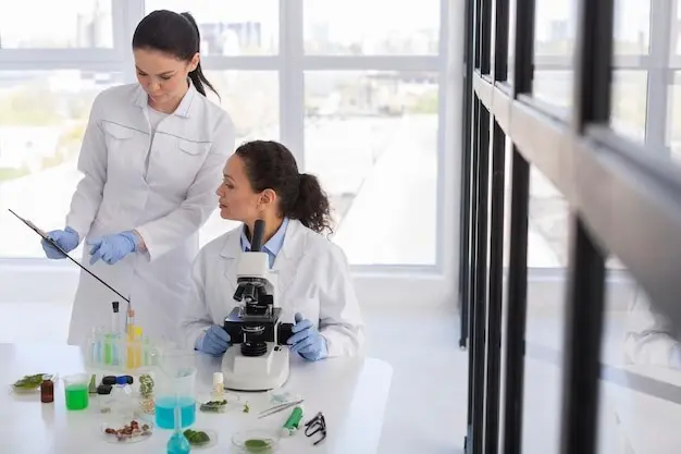
Constructing Individual Models
Personal models begin with structure from anatomy and function from physiology. Segmented MRI or CT outlines geometry, while echocardiography, spirometry, or metabolic panels inform dynamics. Electronic records contribute comorbidities, medications, and trajectories. The pipeline harmonizes units, resolves conflicting measurements, and initializes states consistently. Iterative calibration and cross-checks produce a stable baseline ready to run patient-specific what-if scenarios without guesswork masquerading as certainty.
Story: Preempting a Dangerous Arrhythmia
A middle-aged runner reported palpitations during recovery. Standard tests were inconclusive, yet the individualized electrophysiology model, tuned with wearable ECG and imaging, predicted reentry under dehydration and high catecholamine load. Hydration protocol adjustments and beta-blocker timing were simulated before implementation. Weeks later, training resumed without events. The outcome was not magic, but disciplined modeling plus data that clarified triggers and guided safer routines.

Continuous Sensing Meets Physiology
When AI and Mechanism Collaborate

Safety, Fairness, and Accountability
Building and Scaling the Ecosystem

All Rights Reserved.